Development of the vascular system
Learning Outcome 1: Formation of the Arterial System from the Aortic Arches
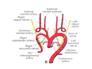
There are six pairs of aortic arches that form in the embryo, arising from the aortic sac. As the embryo develops, these arches undergo a series of transformations, with some disappearing and others maturing into various adult arterial structures. The derivatives of each arch include:
- First aortic arch: Contributes to the formation of the maxillary artery.
- Second aortic arch: Develops into the stapedial artery and hyoid artery.
- Third aortic arch: Gives rise to the common carotid artery and the proximal part of the internal carotid artery.
- Fourth aortic arch: Forms the arch of the aorta (left side) and the proximal part of the right subclavian artery (right side).
- Fifth aortic arch: Typically regresses and does not contribute significantly to the adult arterial system.
- Sixth aortic arch: Becomes the pulmonary arteries and the ductus arteriosus (left side).
Learning Outcome 2: Formation of the Venous System from the Sinus Venosus
The sinus venosus is an embryonic structure that receives blood from the vitelline, umbilical, and cardinal veins. During development, the sinus venosus undergoes remodeling to form the major veins in the adult venous system. Key transformations include:
- The right horn of the sinus venosus develops into the smooth part of the right atrium (sinus venarum) and the sinoatrial node.
- The left horn of the sinus venosus regresses, leaving only a small remnant called the oblique vein of the left atrium.
- The vitelline veins form the portal venous system, including the hepatic portal vein.
- The umbilical veins contribute to the formation of the ligamentum teres (round ligament) of the liver and the ductus venosus.
- The cardinal veins undergo remodeling to form the superior and inferior vena cava, as well as the azygos and hemiazygos veins.
Learning Outcome 3: Congenital Anomalies and Clinical Importance
The congenital anomalies associated with the development of the arterial and venous systems; Some common anomalies and their clinical implications include:
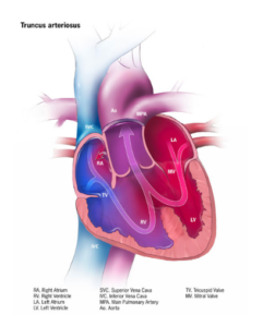
- Persistent truncus arteriosus: A single arterial trunk arises from the heart, instead of separate aorta and pulmonary arteries. This condition results in a mixing of oxygenated and deoxygenated blood, leading to cyanosis and heart failure.
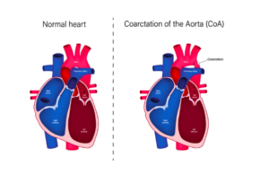
- Coarctation of the aorta: A narrowing of the aortic arch or descending aorta, which can cause hypertension in the upper body and decreased perfusion in the lower body.
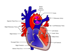
- Patent ductus arteriosus: Failure of the ductus arteriosus to close after birth, resulting in a left-to-right shunt that can lead to heart failure and pulmonary hypertension.
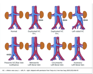
- Inferior vena cava anomalies: Anomalies such as duplicated, interrupted, or left-sided inferior vena cava can have implications for surgical and interventional procedures, as well as increase the risk of deep vein thrombosis.